Gastrointestinal endoscopy - gold standard for early diagnosis gastrointestinal cancer
Gastrointestinal endoscopy is a direct examination method that helps to accurately detect lesions in the digestive tract, while other methods can not, especially when a lesion is at early stage.
What is gastrointestinal endoscopy?
Gastrointestinal endoscopy is the common name for gastroscopy and colonoscopy. This is one of the most modern methods available help to diagnose accurately and easily many diseases related to the digestive tract. In particular, this method have great significance in the early diagnosis of gastrointestinal cancers such as rectal cancer, gastric cancer, duodenal cancer...
To perform gastrointestinal endoscopy, a doctor use a small, lightweight endoscope with a camera and light on the tip. The endoscope will be inserted from the nose or mouth to view the upper digestive tract including: esophagus, stomach, duodenum; or inserted from the anus to observe the lower digestive tract including: colon, rectum, anus.
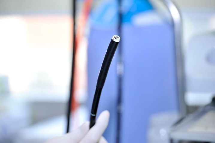
Photo: Endoscope in Hoang Long Clinic
When is an endoscope necessary?
When experiencing the following symptoms, patients should immediately go to a gastroenterology department for examination and producing endoscopy.
- Frequent epigastric pain
- Vomiting blood, rectal bleeding, black stool
- Heartburn, belching, nausea, vomiting
- Sore throat, persistent cough, chest pain, shortness of breath that is not caused by a respiratory disease
- Difficulty swallowing, swallowing as if there is a foreign object stuck in the throat
- Fatigue, loss of appetite, unexplained weight loss
- People who are monitoring digestive diseases such as ulcers, polyps, ..
- Especially those who want to screen for early gastrointestinal cancer or those with risk factors such as: having a relative with gastrointestinal cancer, ...
Popular gastrointestinal endoscopy method
Normal gastrointestinal endoscopy
For this procedure, the endoscope will be inserted through the nose or mouth (upper gastrointestinal endoscopy - gastroscopy) or through the anus (lower gastrointestinal endoscopy -colonoscopy). During the endoscopy, the patient is fully awake and may experience some symptoms such as pain, nausea, vomiting, discomfort, etc.
Anesthesia gastrointestinal endoscopy
The patient will be anesthetized prior to the procedure. The anesthetic process take a short time (about 15-20 minutes) so the patient can wake up immediately after finishing. With this method, the patient will feel light, comfortable and there was no pain during examination.
Magnifying endoscopy – detecting cancer in early stage
If in the past, endoscopy was performed using ordinary white light, many gastrointestinal lesions could be missed. Currently, with advanced endoscopy equipment such as laser endoscopes, magnifying endoscopes, virtual staining technique... will accurately detected even the smallest lesions in the gastrointestinal tract.
Endoscopy Center - Hoang Long Clinic is proud of being the units that have successfully applied 4.0 technology to endoscopic techniques, this help to diagnose and detect accurately gastrointestinal cancer in early stage.
- Endoscope with magnification: The optical and digital magnification of approximately 300 times (equivalent to a microscope) helps to clearly observe the microvascular structure and surface of a lesion, thereby identifying whether it is benign or malignant
- When a lesion is suspected as malignant, in addition to magnification, the doctor will use LCI (Linked Color Imaging) virtual staining to detect early stage cancer – even very early, when cancer cells only exist in the mucosal layer.
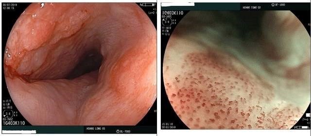
Photo: Early stage Esophagus Cancer detected by endoscopy with ordinary white light (left) and with magnifying and virtual staining technique (right) in Hoang Long Clinic.
- BLI, BLI-Bright technology (using narrowband light) highlights the lesion, combine with magnifying feature to help doctors distinguish various lesions and easily identify the lesion which is benign or potentially malignant.
- Integrated software for early cancer detection and Fuji Intelligent Color Enhancement (FICE) function help identify the change of lesion surface with high-resolution. If a lesion is suspected as cancerous, this lesion will be redder than usual. As a result, it is possible to detect cancer at VERY EARLY stage with high accuracy when compared with biopsy results.
According to statistics, digestive tract cancer such as colon cancer, if detected at an early stage, the survival rate is up to 90%. Many people have been living healthy lives for more than 20 years after being diagnosed with this disease. Therefore, when noticing any abnormal signs, the patient should go to the doctor for timely screening and treatment in order to avoid the serious progression.
Contact information: Hoang Long Clinic
- Address: 10th floor, VCCI tower, 9 Dao Duy Anh, Dong Da, Hanoi.
18th floor, CONINCO tower, 4 Ton That Tung, Dong Da, Hanoi.
- Hotline: 19008904| 024 628 11 331
- Zalo: 0986954448
- Fanpage: https://www.facebook.com/phongkhamdakhoahoanglong/
- Gastrointestinal endoscopy - gold standard for early diagnosis gastrointestinal cancer
- Current methods of small bowel endoscopy
- AMULET Innovality – The most accurate mammography technology
- When to stop treatment for hepatitis B
- Endoscopic ultrasound – an advanced technique for diagnosing gastrointestinal tumors